
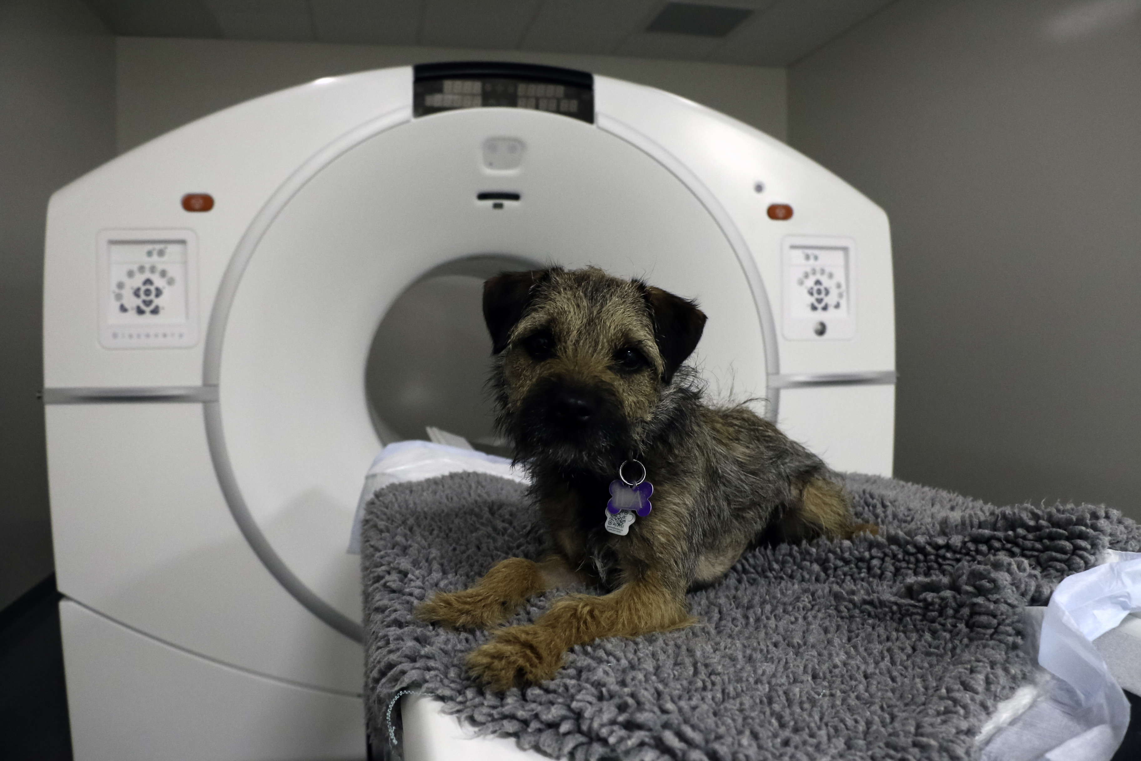
Veterinary college at USask opens Canada’s first PET-CT unit for animals
Western Canada’s regional veterinary college, based on the University of Saskatchewan (USask) campus, is now home to Canada’s only PET-CT unit dedicated to clinical use in animals as well as for animal-human research studies.
By Jeanette Neufeld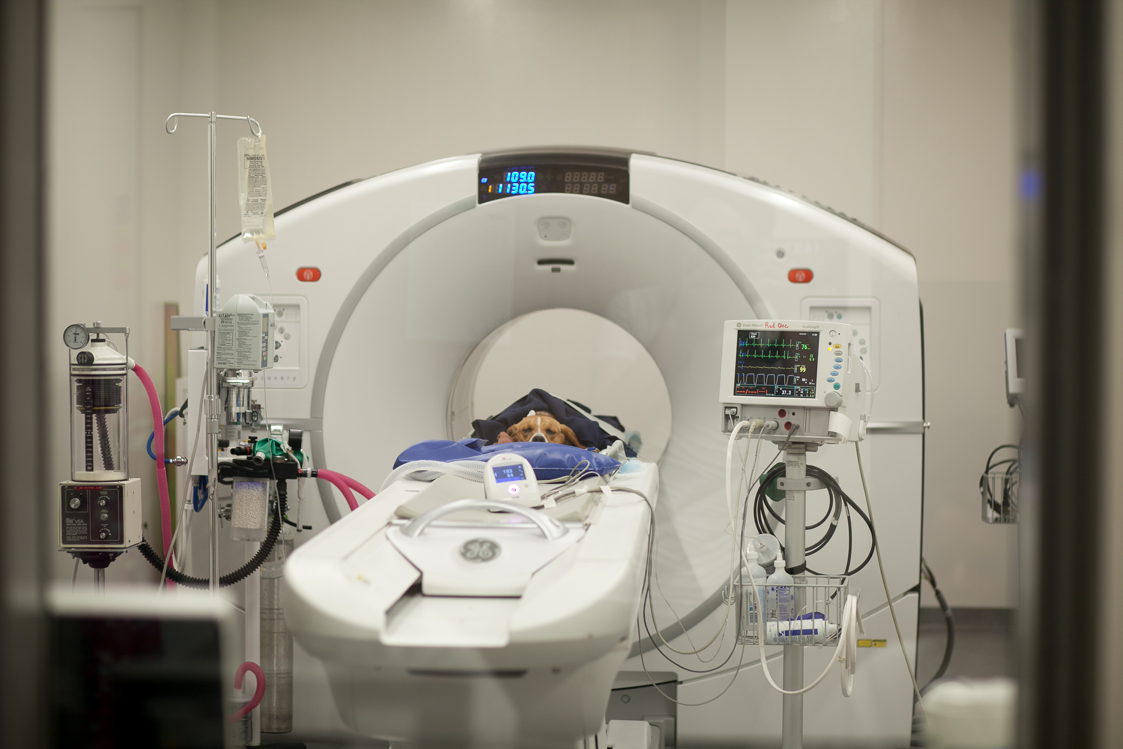
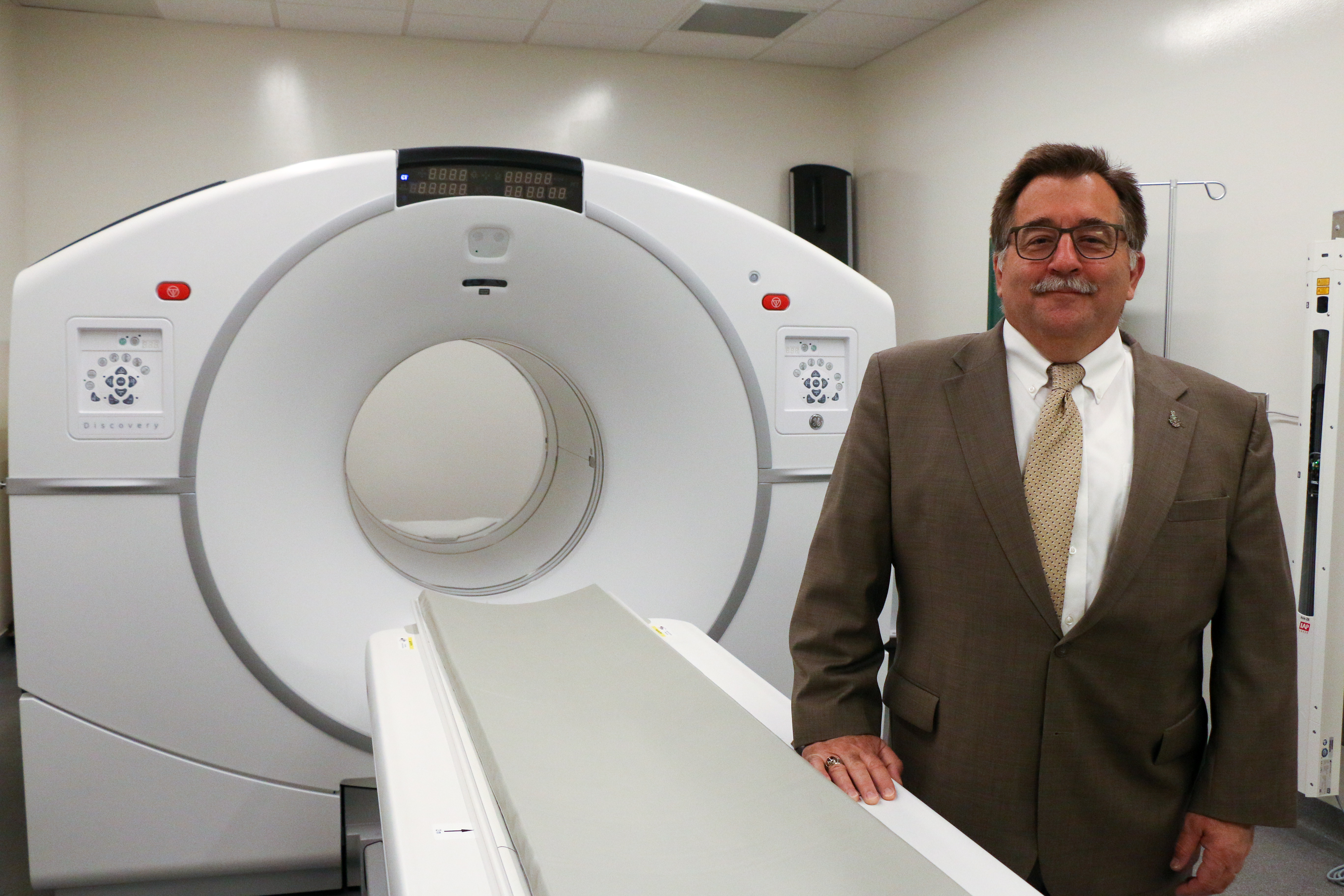
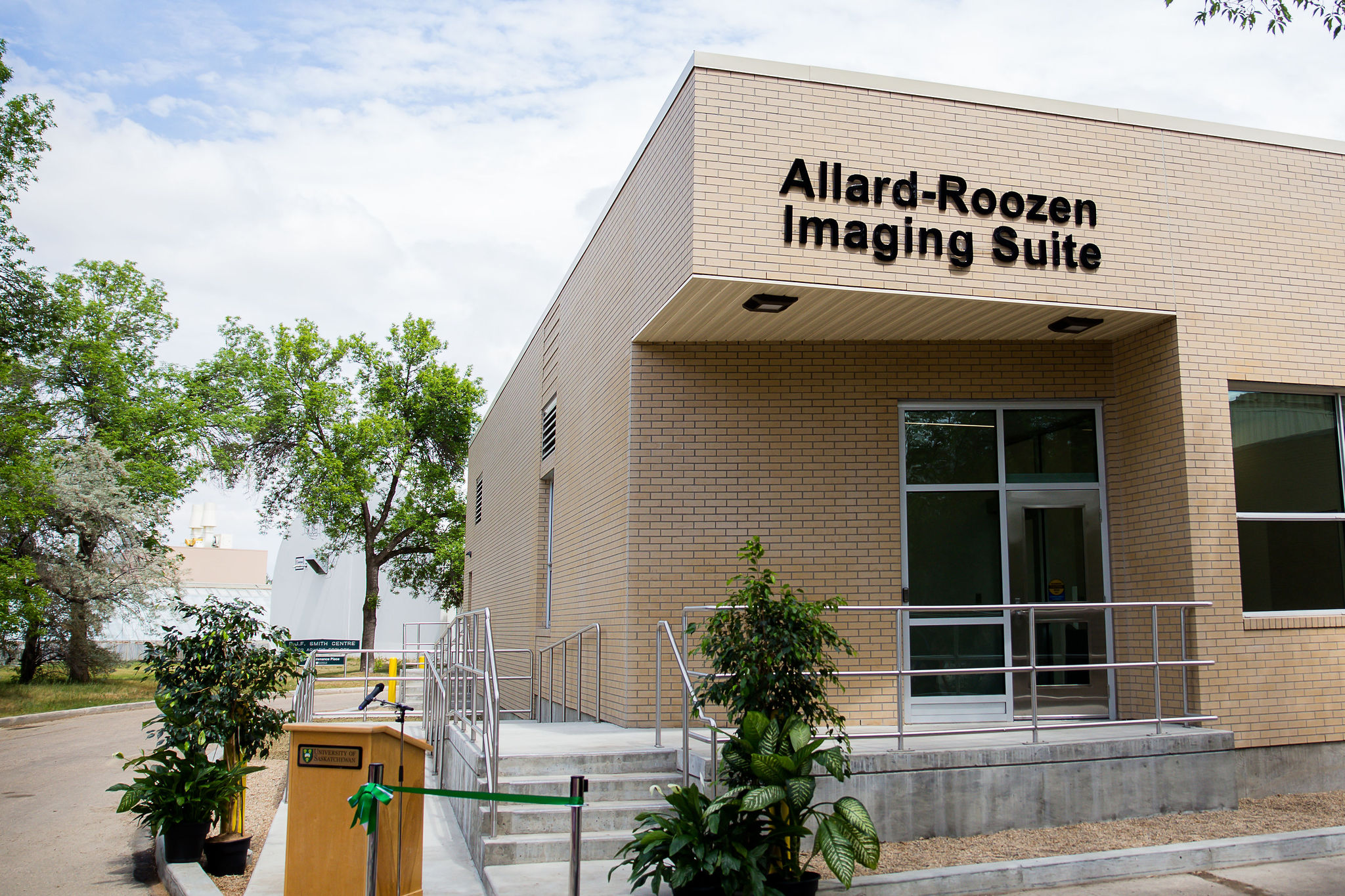
The Western College of Veterinary Medicine (WCVM) celebrated the official opening of the Allard-Roozen Imaging Suite on June 7. The suite’s construction and purchase of the PET-CT scanner was made possible by a $2.5-million donation from Cathy Roozen, an Alberta businesswoman and philanthropist.
“We’re extremely grateful to Cathy for this very generous gift that is allowing this PET-CT suite to finally become a reality,” said Dr. Douglas Freeman, dean of the WCVM. “Her belief in our veterinary college’s ability to accomplish great things in the areas of oncology, medical imaging and One Health is a major motivator for our clinical and research teams.”
With this technology, USask joins a select group: only five other North American universities with veterinary colleges have a PET-CT unit available for clinical use in animals and for research studies.
A positron emission tomography-computed tomography (PET-CT) scan combines the information available from a CT scan – a three-dimensional X-ray – with a PET scan, which delivers information about the metabolic activity in tissues.
For example, PET-CT technology can help detect cancer, brain disorders, heart disease and infections before any anatomical changes are detectable by other imaging scans.
PET-CT technology relies on the use of radioisotopes. The new Allard-Roozen Imaging Suite is located only metres away from the Sylvia Fedoruk Canadian Centre for Nuclear Innovation where the short-lived radioisotopes are produced in the centre’s cyclotron.
“There could be no better location for this new imaging suite,” said Freeman. “An on-site PET-CT scanner opens so many more opportunities for our researchers to collaborate with other scientists across campus and across the country on One Health studies.”
The new imaging suite will enhance the WCVM’s oncology services and support collaborative One Health research studies targeting animal and human health.
A PET-CT scanner increases capabilities of WCVM veterinary specialists to diagnose cancers and to determine their extent. Because cancer cells have a more rapid metabolism than normal cells, a PET scan enables veterinary oncologists to pinpoint abnormal cells much more accurately than they could with standard imaging tools.
Oncology studies in pets can also serve as a model for certain types of cancers that affect people, adding to the importance of these clinical trials.
“Our goal is to allow our patients to live longer, cancer-free lives,” said Dr. Monique Mayer, a specialist in radiation oncology and a professor in the WCVM Department of Small Animal Clinical Sciences. “With the addition of this machine, there are no longer any barriers to the type of treatment we can offer. I am so excited about the possibilities the new Allard-Roozen Imaging Suite opens for our research and our treatment of patients at the WCVM.”
The research potential of the PET-CT machine expands beyond the study of cancer to include a wide array of researchers in different areas of animal and human health, including reproductive science.
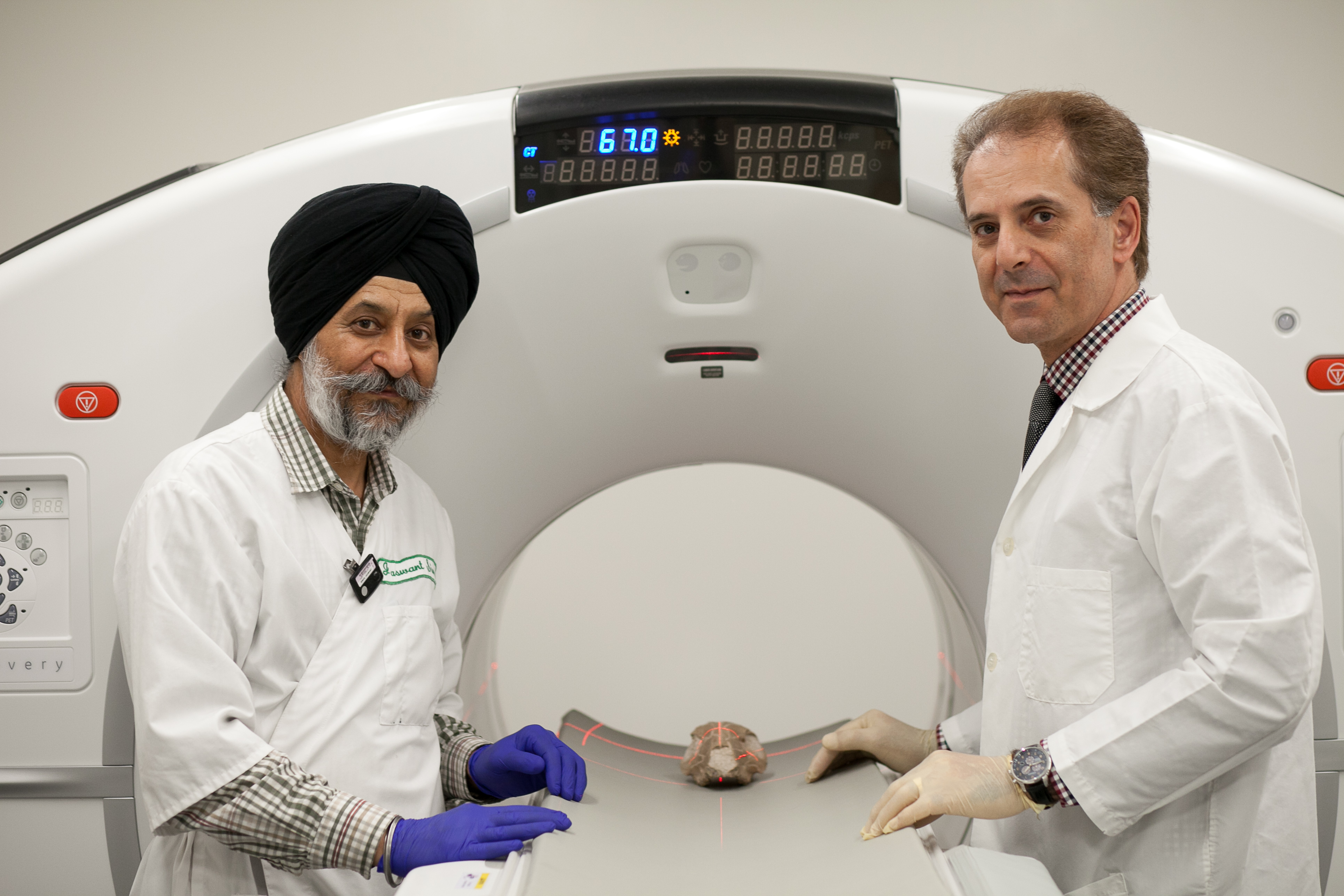
For example, WCVM scientists are developing a technique called “biotracking” that will help them follow the path of certain molecules as they interact in the brains of llamas during induced ovulation. By viewing the movement of a specific chemical in the brain, reproductive physiologists can understand more about how ovulation works in the species, and potentially achieve commercial application for this knowledge.
This technique is only possible through the collaboration of WCVM scientists with researchers at the nearby cyclotron and in other health science colleges across the USask campus.
“We are so proud that we were able to accomplish everything here on campus. It’s taken nearly seven years to put the whole team together,” said Dr. Jaswant Singh, a reproductive physiologist and professor based at WCVM.
He sees potential for boundless collaboration thanks to the addition of the new PET-CT unit at the WCVM.
“This is the centrepiece that allows us to think very broadly and connect all the pieces together,” said Singh.
For more information, contact:
Jeanette Neufeld
Communications Co-ordinator
Western College of Veterinary Medicine
University of Saskatchewan
306-966-2560
jeanette.neufeld@usask.ca